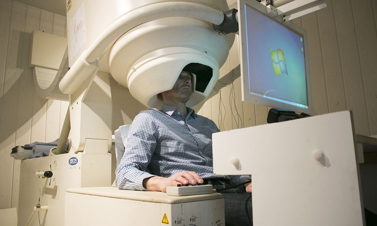
MEG Technology

MagnetoEncephalography (MEG) is a type of scan that measures the magnetic fields generated by brain activity. These extremely small magnetic fields pass through the skull and scalp as if they were transparent, and can therefore be measured outside of the head using the MEG scanner.
When is MEG used?
MEG scans can provide different types of information such as:
- What activity is the brain producing and where in the brain does it come from? For example, it can be used to measure brain activity associated with relaxation, migraine and epilepsy.
- Which part of the brain undertakes different tasks? For example, MEG can determine exactly which part of your brain controls actions such as speaking or moving arms and legs.
- How does the brain work? MEG researchers are working to further understand the way in which the brain functions both normally, and when something goes wrong.
Who can have an MEG scan?
Metallic objects such as cardiac pacemakers, metallic implants or fragments in the eye, and dental implants/wiring interfere with the signal, meaning the data that is collected will not be useful to the researcher. This is not a safety concern since MEG does not use a magnet. Although there is no evidence to suggest that MEG is harmful to unborn babies, as a precaution, the Department of Health advises against scanning pregnant women unless there is a clinical benefit. We do not test for pregnancy as routine so if you think you may be pregnant you should not take part in this study.
What happens during the scan?
Each scanning session will be part of a specific study. The first twenty minutes will be spend setting up the equipment and making sure that you are in a comfortable position in the chair. Blankets are also available if needed. The researcher will then explain in detail exactly what tasks you will have to carry out whilst being scanned. All that we need you to do is to keep still and perform the task to the best of your ability.
Unlike in MRI, there will be no loud noises or buzzing during the scan. Our imaging team members will assist you throughout the procedure and you will be able to talk with them through an intercom at any time. If you feel uncomfortable you can call them just by pressing a buzzer which will be in your hand at all times during the scan.
What are the perks of volunteers undergoing MEG?
When you volunteer to have an MEG scan you leave with the knowledge that the experience has contributed to furthering the progress of medical and psychological research.
What happens after the scan?
After the session has finished we will remove you from the scanner, and the researcher will accompany you to collect your belongings.
Are there any side effects?
There are no know side effects to MEG scanning.
Can I see/receive a picture of my brain?
Depending on the study that you are participating in, it may be possible for you to receive a picture of your brain. Please ask the Researcher for the study that you are taking part in.

MEG is shedding light on some of the fundamental workings of the human brain. Studies that involve normal volunteers form the basis of this and are a vital part of our research program. Through such work, we are learning about normal brain function in areas such as language, vision, movement, memory, thought and emotion.
To find out about studies which involve MRI brain scanning, please see the News section of our website.


